

Neo-IsoCore - our offer
Neo-IsoCore is fully equipped to support tritium (³H)–based studies across a broad range of biomedical research applications, including drug discovery, PET tracer validation, receptor characterization, and molecular imaging.
Whether your research focuses on neurodegenerative diseases, oncology, cardiovascular disorders, endocrine biology, or other translational research areas, Neo-IsoCore offers optimized workflows, advanced imaging capabilities, and expert methodological support to accelerate high impact scientific discovery.
With our facility, you can:
- Screen compound libraries to identify new drug candidates with high affinity for specific molecular targets
- Visualize and quantify target protein distribution across different anatomical regions and tissue types
- Assess target-specificity and selectivity of candidate drugs or PET tracers
- Perform comparative and correlation analyses between multiple imaging tracers
- Validate the pharmacological profiles of novel radioligands prior to in vivo imaging studies
- Map receptor density and distribution in human and animal tissue samples
- Correlate ante-mortem and post-mortem imaging data for target validation and translational insight
- Perform Real-time monitoring of enzyme catalysis
Methods offered
Radioligand binding assays
Radioligand binding is a highly accurate, quantitative, and high-throughput method for studying the pharmacological properties of compounds targeting specific proteins. This method uses tissue or cell homogenates normalized to defined protein concentrations, achieving excellent reproducibility and quantitative precision.
Types of radioligand binding assays
Saturation binding assays
Samples are incubated to equilibrium with increasing concentrations of tritiated radioligand to determine:
- Bmax (maximum receptor density): the total number of binding sites available
- Kd (equilibrium dissociation constant): a measure of binding affinity
Non-specific binding is measured by adding excess unlabeled competitor that fully displaces the radioligand. This assay is fundamental for characterizing new radioligands and understanding target expression levels.
Competition binding assays
Samples are incubated with a fixed concentration of radioligand plus increasing concentrations of unlabeled test compounds. These assays are used for:
- High-throughput screening – Test dozens to hundreds of compounds for target affinity
- Affinity determination – Calculate IC50 and Ki values for candidate drugs
- Pharmacological validation – Confirm that a new radioligand binds the intended target by displacement with known reference compounds
- Selectivity profiling – Test binding to related receptor subtypes or off-target proteins
Regional binding assays
Tissue sections or discrete anatomical regions are incubated with radioligand to assess spatial distribution of binding sites within an organ (typically brain). These assays are used to determine:
- Regional target distribution: Identify where target receptors/proteins are expressed across different anatomical areas
- Relative receptor density: Compare binding levels between regions (e.g., cortical vs. subcortical regions)
- Anatomical selectivity: Evaluate whether a radioligand preferentially binds to specific functional regions.
Autoradiography
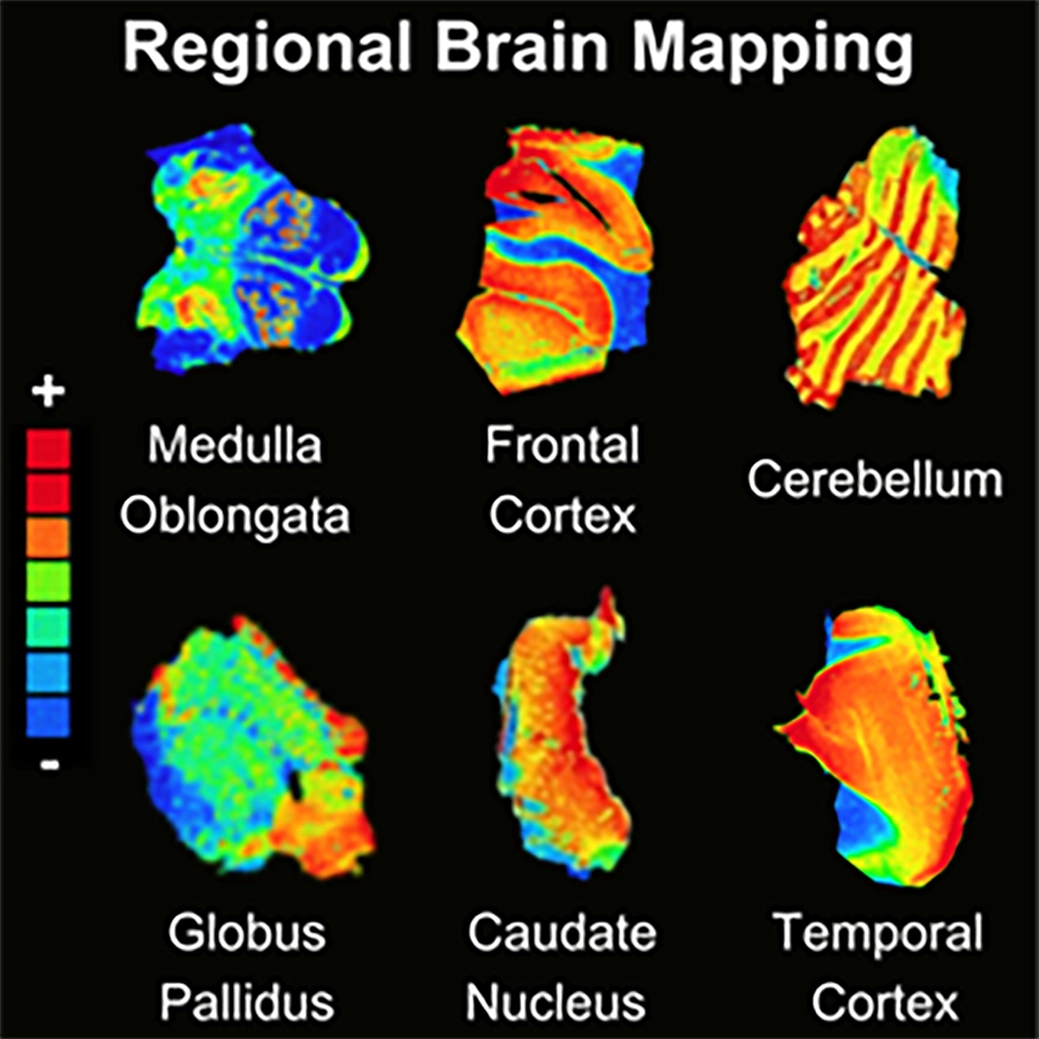
Autoradiography is a quantitative in vitro or ex vivo method used to visualise and quantify the binding of tritium-labeled radioligands to specific molecular targets in tissue sections from post-mortem human tissues or animal models.
This technique is essential for:
- Drug development: Initial characterization of a compound's tissue distribution, regional binding patterns, and target engagement
- PET tracer validation: High-resolution mapping that confirms region-specific and target-specific binding before in vivo imaging studies
- Receptor mapping: Detailed anatomical localization of neurotransmitter receptors, hormone receptors, and other binding sites
- Pathology studies: Detection and quantification of disease-related protein aggregates in tissue from disease models or human donors
Key advantages of autoradiography
- Preserves anatomical context → Visualise exactly regions or brain layers where binding occurs within complex tissue architecture
- High spatial resolution → Our BAS-2500 scanner achieves up to 50 μm pixel resolution
- Quantitative measurement → Correlate binding signal intensity with target density
- Multi-region analysis → Compare binding across brain regions, tumor types, or disease-affected tissues simultaneously
Sample types we work with
- Tissue homogenates from animal models
- Post-mortem human brain tissue
- Membrane preparations enriched for target proteins
- Fresh or frozen tissue samples
Instruments

Tri-Carb 2910TR PerkinElmer
Large-volume liquid scintillation beta counter.
- Optimized for 5-25 ml sample tubes
- Sample capacity 408 standard 20 ml vials, or 720 small 5 ml vials.
- Quick-Count sample loading
This instrument is ideal for:
- Tissue homogenate binding assays with larger sample volumes
- Tritium detection with high sensitivity and wide dynamic range
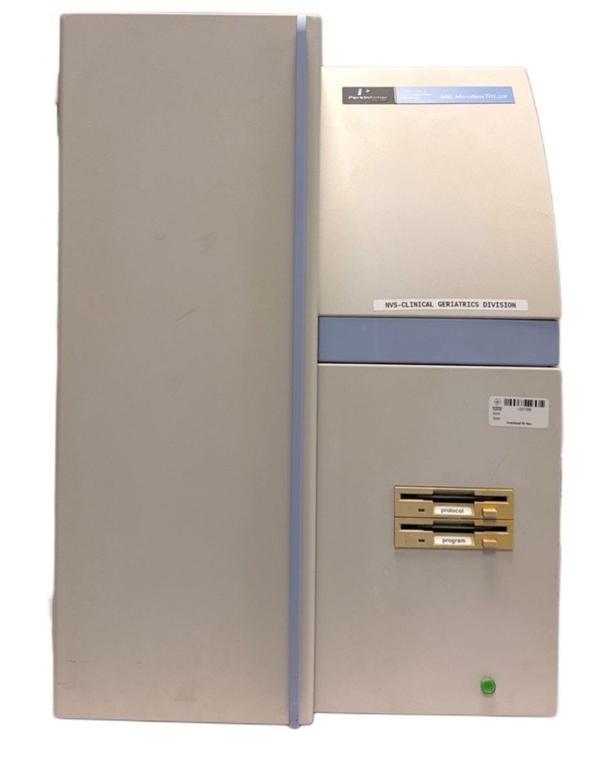
MicroBeta Trilux 1450
High-throughput LSC and luminescence counter (96-well format).
Key features:
- 96-well plate compatibility for high-throughput analysis
- Simultaneous detection of beta radiation and luminescence signals
- High sensitivity for low-level tritium detection in small-volume samples
- Low background noise and high signal-to-noise ratio
This instrument is ideal for:
- High-throughput screening of compounds in competition binding assays
- Rapid quantification of radioligand binding
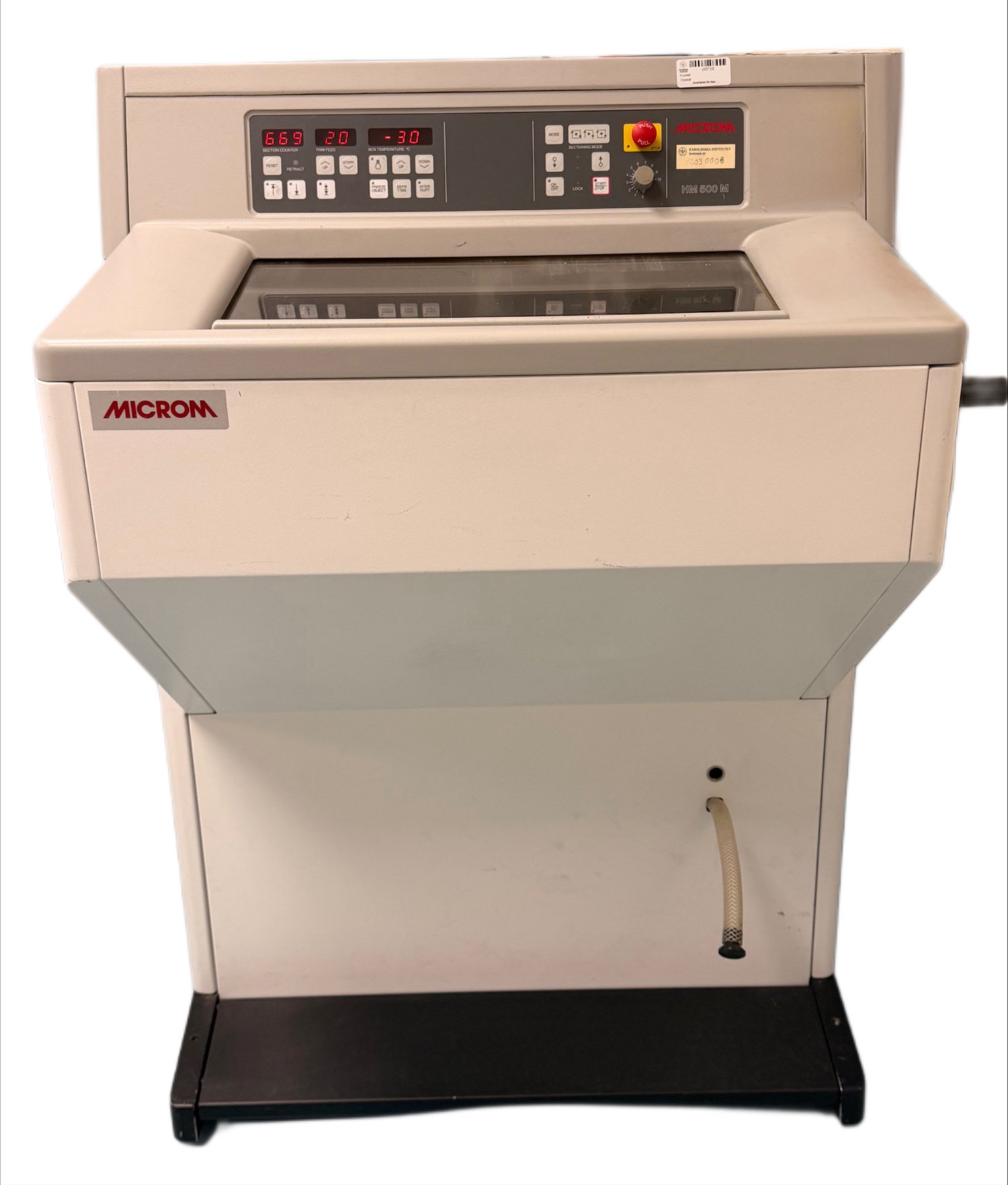
Microm HM 500 & 560M Cryostat
Used for sectioning frozen tissue.
Precision cryomicrotome for routine tissue sectioning with:
- Section thickness range: 1-60 μm
- Stable temperature control for optimal cutting of frozen tissue
- Large specimen capacity for diverse tissue types
- Low-noise cooling system
- Fast freezing station
- Single, interval and continuous stroke operating modes
- Integrated swiveling knife guard
- Universal micro/macro feed system
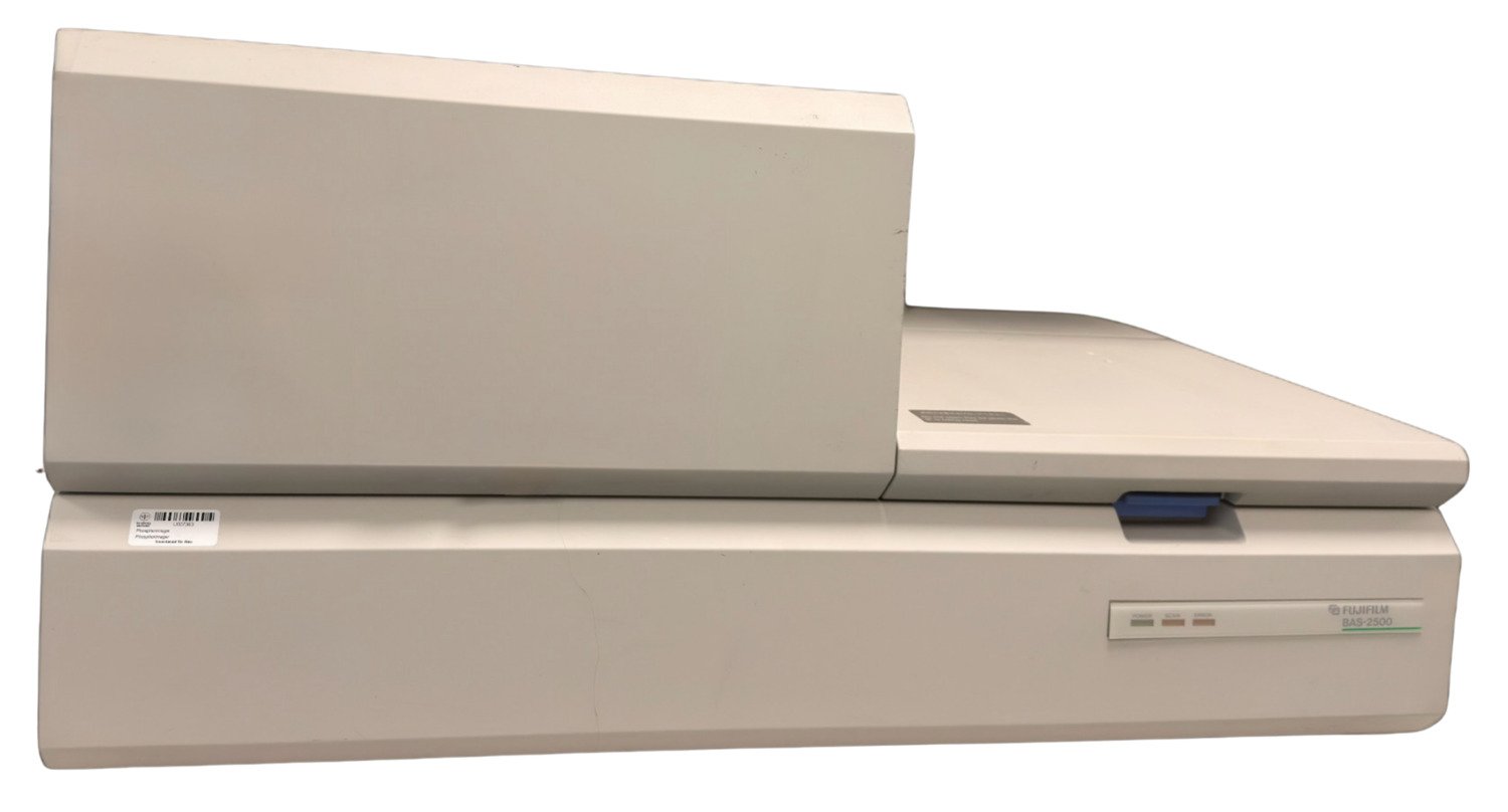
Fujifilm BAS-2500 Scanner
Used for section-based imaging and quantification of radioligand binding in tissue sections.
Phosphor imaging plate scanner for quantitative autoradiography with:
- Pixel resolution up to 50 μm for high-detail anatomical mapping
- 20x40 cm imaging plate can be scanned within five minutes at 16 bits
- Wide dynamic range for simultaneous detection of high and low binding regions
- Quantitative analysis using calibrated tritium standards
- IP Size: 20x40 cm
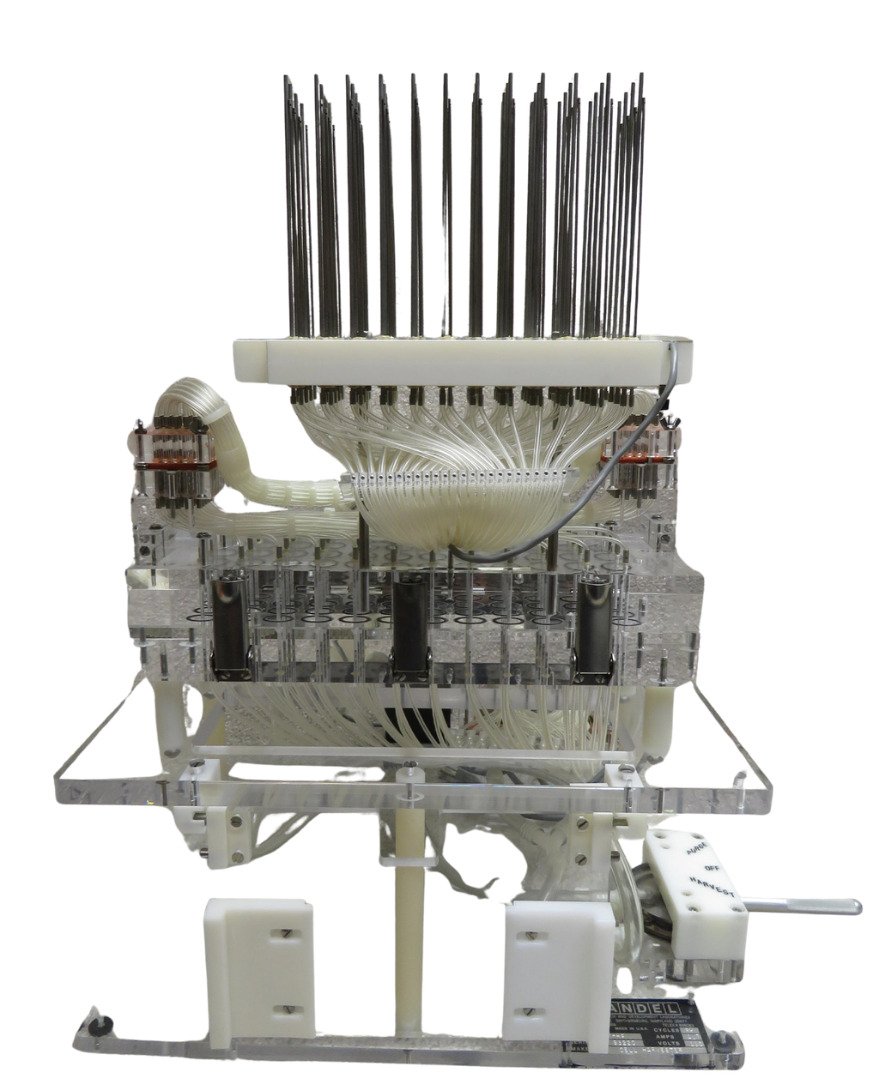
Conventional sample harvester
Used for filtration-based separation of bound and free radioligand in large sample assays.
Manual/semiautomated harvesting system for radioligand binding assays.
For large sample radioligand binding assays.
- Vacuum filtration through glass fiber filters
- Suitable for large numbers of tube-based samples
- Compatible with standard binding assay formats
- Robust and reliable for routine use

PerkinElmer FilterMate Harvester
Automated 96-well format harvesting.
Used for high-throughput filtration in plate-based radioligand binding assays
Key features:
- Automated vacuum filtration for 96-well plates
- Rapid and reproducible separation of bound/free radioligand
- Reduced hands-on time and improved assay consistency
- Compatible with filter plates and scintillation counting workflows
- Designed for integration with high-throughput screening systems
Lab bench
Designated laboratory workspace for safe handling of radioactive materials and BSL2 work.
Used for preparation, handling, and disposal of radiolabeled compounds under controlled and compliant conditions.
- Certified for work with radioactive isotopes (e.g., tritium)
- Compliance with radiation safety and laboratory regulations
- Radioactive waste storage and disposal vis Radwaste online system registration
This setup is ideal for:
- Preparation of radioligand solutions
- Handling and dilution of radioactive compounds
- Safe transfer of samples during binding assays
- Minimizing contamination and ensuring operator safety
Safety, Storage and Waste disposable Infrastructure
- Designated cold storage units for safe and compliant handling of radioactive materials
- Used for storage and preservation of radiolabeled compounds under controlled temperature conditions
- Certified for use with radioactive substances in accordance with KI radiation safety regulations
- Certified for radioactive waste storage, disposal and Radwaste registration
- Compliance with laboratory and radiation protection guidelines
This setup is ideal for:
- Storage of radioligands and isotope-labeled reagents at -20 °C
- Short-term storage of radioactive samples in refrigerators (2–8 °C)
- Maintaining stability and integrity of temperature-sensitive compounds
- Segregation of radioactive materials from non-radioactive laboratory items
- Ensuring safe, traceable, and regulated storage of isotopes
- Safe storage and disposal of radioactive waste and material
Research applications
Ex Vivo radioligand binding studies
- Competition binding assays (IC50, Ki determination)
- Saturation binding assays (Bmax, Kd quantification)
- Regional receptor mapping in tissue sections
- Drug candidate screening and validation
Autoradiography and imaging
- Quantitative receptor autoradiography
- Spatial distribution mapping of molecular targets
- Human and animal tissue analysis
- Pre-clinical PET tracer validation
Potential research areas we can support
Drug Discovery & Pharmacology
- Quantitative receptor binding assays (Bmax, Kd)
- Ligand screening and pharmacological profiling
- Kinetic characterization of receptor–ligand interactions
- Applications in GPCR-targeted drug discovery, allosteric modulation, and theranostic radioligand development
Neurobiology & Neurodegeneration
- Spatial mapping of receptor distribution in postmortem brain
- Quantification of pathological protein aggregates
- Preclinical validation of PET radiotracers
Molecular Imaging & Radiopharmaceuticals
- Development and validation of PET radiotracers
- Quantitative correlation of in vitro binding data with in vivo imaging outcomes
- Applications in CNS and oncology imaging
Oncology
- Quantitative tumor receptor profiling
- Development and characterization of targeted radioligand therapies
- Applications include somatostatin receptor imaging and PSMA-targeted agents
- Real-time enzymatic catalysis monitoring
Cardiovascular & Endocrine Research
- Characterization of hormone–receptor interactions
- Functional and binding studies of GPCRs in tissue systems
Other Translational Aspects
- In vitro–in vivo correlation
- Target identification & quantification
- Mapping of spatial and temporal distribution of molecular targets